Chemical Biology

Functional Metabolomics and Lipidomics

Functional Metabolomics and Lipidomics image 
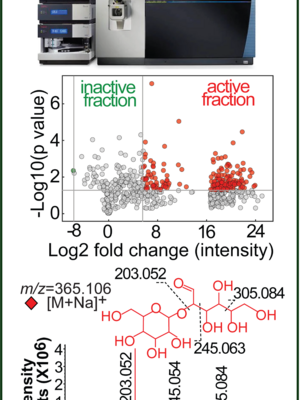
We are establishing metabolomic and lipidomic platforms to profile and identify various metabolites and lipids that bind and interact with antigen-presentation molecules and other proteins. Metabolites can be defined as polar or non-polar small molecules with mass units generally less than 2000 and as direct products of various metabolic pathways (human metabolome database). More narrowly, water-soluble metabolites are sometimes designated “metabolites” to separate them from lipids. Small lipids and metabolites are critical compounds derived from various metabolic pathways to control and regulate immune pathways through binding and interacting with antigen-presentation molecules, transcription factors, hormone receptors, and other proteins. We will set up the metabolomic capability to profile and identify biologically important compounds in immune responses, microbial infections, and cancers. We will also integrate bioassay-guided isolation, serial or 2D high-performance liquid chromatography, and other chemical and biochemistry techniques for compound preparation and identification.

Representative Publications

  • Huang, Shouxiong, Adam Shahine, Tan-Yun Cheng, Yi-Ling Chen, Soo Weei Ng, Gautham R Balaji, Rachel Farquhar, et al. (2023) 2023. “CD1 Lipidomes Reveal Lipid-Binding Motifs and Size-Based Antigen-Display Mechanisms”. Cell 186 (21): 4583-4596.e13. https://doi.org/10.1016/j.cell.2023.08.022.

    The CD1 system binds lipid antigens for display to T cells. Here, we solved lipidomes for the four human CD1 antigen-presenting molecules, providing a map of self-lipid display. Answering a basic question, the detection of >2,000 CD1-lipid complexes demonstrates broad presentation of self-sphingolipids and phospholipids. Whereas peptide antigens are chemically processed, many lipids are presented in an unaltered form. However, each type of CD1 protein differentially edits the self-lipidome to show distinct capture motifs based on lipid length and chemical composition, suggesting general antigen display mechanisms. For CD1a and CD1d, lipid size matches the CD1 cleft volume. CD1c cleft size is more variable, and CD1b is the outlier, where ligands and clefts show an extreme size mismatch that is explained by uniformly seating two small lipids in one cleft. Furthermore, the list of compounds that comprise the integrated CD1 lipidome supports the ongoing discovery of lipid blockers and antigens for T cells.

  • Huang, Shouxiong, Tan-Yun Cheng, David C Young, Emilie Layre, Cressida A Madigan, John Shires, Vincenzo Cerundolo, John D Altman, and Branch Moody. (2011) 2011. “Discovery of Deoxyceramides and Diacylglycerols As CD1b Scaffold Lipids Among Diverse Groove-Blocking Lipids of the Human CD1 System”. Proceedings of the National Academy of Sciences of the United States of America 108 (48): 19335-40. https://doi.org/10.1073/pnas.1112969108.

    Unlike the dominant role of one class II invariant chain peptide (CLIP) in blocking MHC class II, comparative lipidomics analysis shows that human cluster of differentiation (CD) proteins CD1a, CD1b, CD1c, and CD1d bind lipids corresponding to hundreds of diverse accurate mass retention time values. Although most ions were observed in association with several CD1 proteins, ligands binding selectively to one CD1 isoform allowed the study of how differing antigen-binding grooves influence lipid capture. Although the CD1b groove is distinguished by its unusually large volume (2,200 Å(3)) and the T' tunnel, the average mass of compounds eluted from CD1b was similar to that of lipids from CD1 proteins with smaller grooves. Elution of small ligands from the large CD1b groove might be explained if two small lipids bind simultaneously in the groove. Crystal structures indicate that all CD1 proteins can capture one antigen with its hydrophilic head group exposed for T-cell recognition, but CD1b structures show scaffold lipids seated below the antigen. We found that ligands selectively associated with CD1b lacked the hydrophilic head group that is generally needed for antigen recognition but interferes with scaffold function. Furthermore, we identified the scaffolds as deoxyceramides and diacylglycerols and directly demonstrate a function in augmenting presentation of a small glycolipid antigen to T cells. Thus, unlike MHC class II, CD1 proteins capture highly diverse ligands in the secretory pathway. CD1b has a mechanism for presenting either two small or one large lipid, allowing presentation of antigens with an unusually broad range of chain lengths.

Metabolites and Lipids as T cell Antigens and Protein Ligands

Metabolites and Lipids as T cell Antigens and Protein Ligands Image 
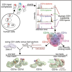
Antigen-presenting molecules bind peptide, lipid, or metabolite ligands to interact with T cell receptor (TCR) and activate different T cell populations. Antigen-presenting molecules, including MR1 and CD1, bind metabolite and lipid ligands to interact with MAIT cells and CD1-restricted T cells, respectively. To identify the metabolites and lipids for T cell activation, we expressed and isolated non-peptidic ligands from MR1 and CD1 proteins to characterize their structures and functions for T cell activation. Taking the CD1 system as an example, human CD1a, CD1b, CD1c, and CD1d proteins bind lipid antigens for surface display to T cells. We solved lipidomes interacting with these four human CD1 proteins and provided a map of self-lipid display via detecting >2,000 CD1-lipid complexes and demonstrating a broad presentation of self-sphingolipids and phospholipids. These data allowed further size-matching of lipids with CD1 lipid-binding clefts and provided candidate lipid blockers and agonists for T cells.

Representative Publications

  • Huang, Shouxiong, Adam Shahine, Tan-Yun Cheng, Yi-Ling Chen, Soo Weei Ng, Gautham R Balaji, Rachel Farquhar, et al. (2023) 2023. “CD1 Lipidomes Reveal Lipid-Binding Motifs and Size-Based Antigen-Display Mechanisms”. Cell 186 (21): 4583-4596.e13. https://doi.org/10.1016/j.cell.2023.08.022.

    The CD1 system binds lipid antigens for display to T cells. Here, we solved lipidomes for the four human CD1 antigen-presenting molecules, providing a map of self-lipid display. Answering a basic question, the detection of >2,000 CD1-lipid complexes demonstrates broad presentation of self-sphingolipids and phospholipids. Whereas peptide antigens are chemically processed, many lipids are presented in an unaltered form. However, each type of CD1 protein differentially edits the self-lipidome to show distinct capture motifs based on lipid length and chemical composition, suggesting general antigen display mechanisms. For CD1a and CD1d, lipid size matches the CD1 cleft volume. CD1c cleft size is more variable, and CD1b is the outlier, where ligands and clefts show an extreme size mismatch that is explained by uniformly seating two small lipids in one cleft. Furthermore, the list of compounds that comprise the integrated CD1 lipidome supports the ongoing discovery of lipid blockers and antigens for T cells.