Immunology

Biology of Metabolite Antigen Presentation

Biology of Metabolite Antigen Presentation Image 
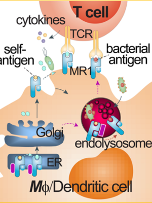
Conventional CD4+ and CD8+ T cells are activated by peptide antigens presented by major histocompatibility complex (MHC) class I and class II molecules, named human leukocyte antigens (HLA) in humans. Peptide antigen presentation has shaped a classical paradigm in T cell activation and vaccine development. Endogenous peptides (e.g., mammalian or viral peptides) and exogenous peptides (bacterial peptides) can differentially utilize secretory, cross-presentation, and endocytic pathways to be loaded onto HLA proteins and activate conventional CD4+ and CD8+ T cells. As unconventional T cells, mucosal-associated invariant T (MAIT) cells and lipid-reactive T cells respond to metabolite and lipid antigens presented by MHC class I-like molecules MHC-related protein 1 (MR1) and CD1, respectively. The non-peptidic metabolite or lipid antigen presentation can involve different sets of accessory proteins and alternative pathways for T cell activation. Our lab will characterize metabolite antigen presentation pathways for MAIT cell activation.

Representative Publications

  • Huang, Shouxiong, Susan Gilfillan, Sojung Kim, Bruce Thompson, Xiaoli Wang, Andrea J Sant, Daved H Fremont, Olivier Lantz, and Ted H Hansen. (2008) 2008. “MR1 Uses an Endocytic Pathway to Activate Mucosal-Associated Invariant T Cells”. The Journal of Experimental Medicine 205 (5): 1201-11. https://doi.org/10.1084/jem.20072579.

    Like CD1d-restricted iNKT cells, mucosal-associated invariant T cells (MAITs) are "innate" T cells that express a canonical TCRalpha chain, have a memory phenotype, and rapidly secrete cytokines upon TCR ligation. Unlike iNKT cells, MAIT cells require the class Ib molecule MHC-related protein I (MR1), B cells, and gut flora for development and/or expansion, and they preferentially reside in the gut lamina propria. Evidence strongly suggests that MAIT cell activation is ligand-dependent, but the nature of MR1 ligand is unknown. In this study, we define a mechanism of endogenous antigen presentation by MR1 to MAIT cells. MAIT cell activation was dependent neither on a proteasome-processed ligand nor on the chaperoning by the MHC class I peptide loading complex. However, MAIT cell activation was enhanced by overexpression of MHC class II chaperones Ii and DM and was strikingly diminished by silencing endogenous Ii. Furthermore, inhibiting the acidification of the endocytic compartments reduced MR1 surface expression and ablated MAIT cell activation. The importance of the late endosome for MR1 antigen presentation was further corroborated by the localization of MR1 molecules in the multivesicular endosomes. These findings demonstrate that MR1 traffics through endocytic compartments, thereby allowing MAIT cells to sample both endocytosed and endogenous antigens.

Biology of Unconventional T cell Responses

Unconventional T cells, particularly MAIT cells, bear innate-like features of activation and responses. MAIT cells can respond to microbial infections in much rapid kinetics through recognizing conserved metabolite antigens presented by MR1 protein. This activation mechanism is independent of individual HLA genetic variability in humans. The relatively high frequency of MAIT cells in various tissue sites in humans provides an ideal model and supports the significance of understanding the roles of MAIT cells in diseases and interventions. We are characterizing the unique immune responses of MAIT cells and other unconventional T cells to understand their functions in infectious diseases, particularly tuberculosis and comorbidities, and in some cancers. Immunology assays, transcriptomics, targeted gene mutation, and animal vs. human tissue models with chemical intervention will be used in our studies.

Diagrams - RESPONDING KINETICS OF MUCOSAL-ASSOCIATED INVARIANT T (MAIT) AND NATURAL KILLER T (NKT) CELLS.

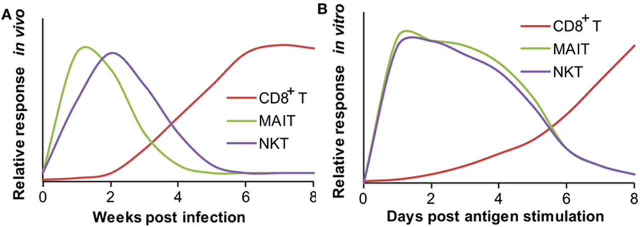
RESPONDING KINETICS OF MUCOSAL-ASSOCIATED INVARIANT T (MAIT) AND NATURAL KILLER T (NKT) CELLS. (A) In vivo responding kinetics were hypothesized based on the presence of mycobacterial antigen-specific CD8+ T cells in lung tissues, the ability of MAIT cells to inhibit Bacillus Calmette–Guérin (BCG) growth in lung tissues, and the ability of transferred iNKT to inhibit M. tuberculosis growth in lung tissues. (B) In vitro-responding kinetics were estimated according to the acquisition of cytolytic function by CD8+ T cells upon in vitro peptide stimulation, cytokine production by polyclonal MAIT cells upon stimulation with BCG-infected macrophages, and cytokine production by tetramer-isolated human polyclonal NKT cells upon antigen-specific activation.

Representative Publications

  • Sharma, Manju, Liang Niu, Xiang Zhang, and Shouxiong Huang. (2023) 2023. “Comparative Transcriptomes Reveal Pro-Survival and Cytotoxic Programs of Mucosal-Associated Invariant T Cells Upon Bacillus Calmette-Guérin Stimulation”. Frontiers in Cellular and Infection Microbiology 13: 1134119. https://doi.org/10.3389/fcimb.2023.1134119.

    Mucosal-associated invariant T (MAIT) cells are protective against tuberculous and non-tuberculous mycobacterial infections with poorly understood mechanisms. Despite an innate-like nature, MAIT cell responses remain heterogeneous in bacterial infections. To comprehensively characterize MAIT activation programs responding to different bacteria, we stimulated MAIT cells with E. coli to compare with Bacillus Calmette-Guérin (BCG), which remains the only licensed vaccine and a feasible tool for investigating anti-mycobacterial immunity in humans. Upon sequencing mRNA from the activated and inactivated CD8+ MAIT cells, results demonstrated the altered MAIT cell gene profiles by each bacterium with upregulated expression of activation markers, transcription factors, cytokines, and cytolytic mediators crucial in anti-mycobacterial responses. Compared with E. coli, BCG altered more MAIT cell genes to enhance cell survival and cytolysis. Flow cytometry analyses similarly displayed a more upregulated protein expression of B-cell lymphoma 2 and T-box transcription factor Eomesodermin in BCG compared to E.coli stimulations. Thus, the transcriptomic program and protein expression of MAIT cells together displayed enhanced pro-survival and cytotoxic programs in response to BCG stimulation, supporting BCG induces cell-mediated effector responses of MAIT cells to fight mycobacterial infections.

  • Sharma, Manju, Shuangmin Zhang, Liang Niu, David M Lewinsohn, Xiang Zhang, and Shouxiong Huang. (2020) 2020. “Mucosal-Associated Invariant T Cells Develop an Innate-Like Transcriptomic Program in Anti-Mycobacterial Responses”. Frontiers in Immunology 11: 1136. https://doi.org/10.3389/fimmu.2020.01136.

    Conventional T cells exhibit a delayed response to the initial priming of peptide antigens presented by major histocompatibility complex (MHC) proteins. Unlike conventional T cells, mucosal-associated invariant T (MAIT) cells quickly respond to non-peptidic metabolite antigens presented by MHC-related protein 1 (MR1). To elucidate the MR1-dependent activation program of MAIT cells in response to mycobacterial infections, we determined the surface markers, transcriptomic profiles, and effector responses of activated human MAIT cells. Results revealed that mycobacterial-incubated antigen-presenting cells stimulated abundant human CD8+ MAIT cells to upregulate the co-expression of CD69 and CD26, as a combinatorial activation marker. Further transcriptomic analyses demonstrated that CD69+CD26++ CD8+MAIT cells highly expressed numerous genes for mediating anti-mycobacterial immune responses, including pro-inflammatory cytokines, cytolytic molecules, NK cell receptors, and transcription factors, in contrast to inactivated counterparts CD69+/-CD26+/- CD8+MAIT cells. Gene co-expression, enrichment, and pathway analyses yielded high statistical significance to strongly support that activated CD8+ MAIT cells shared gene expression and numerous pathways with NK and CD8+ T cells in activation, cytokine production, cytokine signaling, and effector functions. Flow cytometry detected that activated CD8+MAIT cells produced TNFα, IFNγ, and granulysin to inhibit mycobacterial growth and fight mycobacterial infection. Together, results strongly support that the combinatorial activation marker CD69+CD26++ labels the activated CD8+MAIT cells that develop an innate-like activation program in anti-mycobacterial immune responses. We speculate that the rapid production of anti-mycobacterial effector molecules facilitates MAIT cells to fight early mycobacterial infection in humans.