Cell Death Pathways

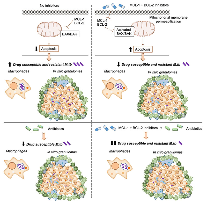
Alveolar macrophages (AMs) are sentinels in the lung which clear inhaled agents, including allergens and microbes; yet do not efficiently clear M. tuberculosis (M.tb). We found that a transcription factor highly expressed in AMs (PPARγ) that is critical for M.tb growth inside human macrophages, induces expression of MCL-1 (family member of anti-apoptotic BCL-2 proteins. Further, MCL-1 limit apoptosis and contributes to M.tb ability to grow intracellularly in human macrophages (Arnett et al. PLoS Pathog 2018). Apoptosis, whereby cells die with intact membranes, limits inflammatory responses and contributes to antigen presentation and T cell activation during M.tb infection (Arnett and Schlesinger Immunity 2021). We are investigating MCL-1 and other anti-apoptotic BCL-2 proteins as targets for host-directed therapy and have found that combinations of inhibitors that are FDA-approved and in clinical trials for cancer therapy significantly reduces growth of drug susceptible and resistant M.tb in human macrophages and a pre-clinical human granuloma model. Since these inhibitors are efficacious in rodent cancer models, and safety and activity studies are underway in cancer clinics, we expect that re-purposing them for TB treatment should translate more readily and rapidly to the clinic than generation of new compounds. Thus, this host-directed therapy approach could signify a breakthrough for TB treatment.

  • Arnett, Eusondia, Ashlee M Weaver, Kiersten C Woodyard, Maria J Montoya, Michael Li, Ky Hoang V, Andrew Hayhurst, Abul K Azad, and Larry S Schlesinger. (2018) 2018. “PPARγ Is Critical for Mycobacterium Tuberculosis Induction of Mcl-1 and Limitation of Human Macrophage Apoptosis.”. PLoS Pathogens 14 (6): e1007100. https://doi.org/10.1371/journal.ppat.1007100.

    Peroxisome proliferator-activated receptor (PPAR)γ is a global transcriptional regulator associated with anti-inflammatory actions. It is highly expressed in alveolar macrophages (AMs), which are unable to clear the intracellular pathogen Mycobacterium tuberculosis (M.tb). Although M.tb infection induces PPARγ in human macrophages, which contributes to M.tb growth, the mechanisms underlying this are largely unknown. We undertook NanoString gene expression analysis to identify novel PPARγ effectors that condition macrophages to be more susceptible to M.tb infection. This revealed several genes that are differentially regulated in response to PPARγ silencing during M.tb infection, including the Bcl-2 family members Bax (pro-apoptotic) and Mcl-1 (pro-survival). Apoptosis is an important defense mechanism that prevents the growth of intracellular microbes, including M.tb, but is limited by virulent M.tb. This suggested that M.tb differentially regulates Mcl-1 and Bax expression through PPARγ to limit apoptosis. In support of this, gene and protein expression analysis revealed that Mcl-1 expression is driven by PPARγ during M.tb infection in human macrophages. Further, 15-lipoxygenase (15-LOX) is critical for PPARγ activity and Mcl-1 expression. We also determined that PPARγ and 15-LOX regulate macrophage apoptosis during M.tb infection, and that pre-clinical therapeutics that inhibit Mcl-1 activity significantly limit M.tb intracellular growth in both human macrophages and an in vitro TB granuloma model. In conclusion, identification of the novel PPARγ effector Mcl-1 has determined PPARγ and 15-LOX are critical regulators of apoptosis during M.tb infection and new potential targets for host-directed therapy for M.tb.

  • Arnett, Eusondia, and Larry S Schlesinger. (2021) 2021. “Live and Let Die: TB Control by Enhancing Apoptosis.”. Immunity 54 (8): 1625-27. https://doi.org/10.1016/j.immuni.2021.07.010.

    The impact of cellular apoptosis in controlling M. tuberculosis during tuberculosis (TB) infection remains unresolved. In this issue of Immunity, Stutz et al. provide compelling evidence that apoptosis controls M. tuberculosis infection in vivo and compounds that induce apoptosis limit M. tuberculosis growth in mice.